

Paper Sharing
【International Papers】Spectral Tuning of Hyperbolic Shear Polaritons in Monoclinic Gallium Oxide via Isotopic Substitution
日期:2026-01-16阅读:371
Researchers from the Fritz-Haber-Institut der Max-Planck-Gesellschaft have published a dissertation titled " Spectral Tuning of Hyperbolic Shear Polaritons in Monoclinic Gallium Oxide via Isotopic Substitution " in Advanced Materials.
Background
Phonon polaritons, quasiparticles arising from the hybridization of photons with infrared (IR)-active phonon modes, exhibit low optical losses and thus hold great potential for applications in nanoscale waveguiding, infrared detection, thermal emission, heat conduction, and sub-diffractional imaging. Recent advances in hyperbolic materials have enabled phonon polaritons to achieve deeply sub-diffractional light confinement, while the anisotropy of the crystal structure further enhances the directionality of polariton propagation. In monoclinic crystals, hyperbolic shear phonon polaritons (HShPs) have been observed, exhibiting asymmetric propagation with optical axes misaligned from the conventional unit cell vectors. Although their directionality can be modulated through nanoantenna design, heterostructure stacking, or thin-slab layering, HShPs are still intrinsically confined to narrow spectral bands, typically spanning only tens to a few hundred cm⁻¹. To expand the accessible spectral range, approaches such as atomic superlattices, heterostructure coupling, and ion intercalation have been explored, while isotopic substitution has emerged as a more direct and effective method for spectral tuning. In materials like hexagonal boron nitride (hBN) and α-MoO₃, isotopic purification has been shown to extend polariton propagation and induce spectral red/blue shifts. However, in low-symmetry three-dimensional polar crystals such as β-Ga₂O₃ (bGO), systematic studies of the effects of ¹⁶O → ¹⁸O isotopic substitution on IR-active phonons and HShPs are still lacking. High-purity isotopic bGO provides the possibility of low-loss polariton propagation, and exploring its spectral tuning effects will help broaden the applications of bGO in nanophotonic devices and lay the foundation for polariton technologies in low-symmetry 3D polar crystals.
Abstract
Hyperbolic phonon polaritons - hybridized modes arising from the ultrastrong coupling of infrared light to strongly anisotropic lattice vibrations in uniaxial or biaxial polar crystals - enable to confine light to the nanoscale with low losses and high directionality. In even lower symmetry materials, such as monoclinic -Ga2O3 (bGO), hyperbolic shear polaritons (HShPs) further enhance the directionality. Yet, HShPs are intrinsically supported only within narrow frequency ranges defined by the phonon frequencies of the host material. Here, we report spectral tuning of HShPs in bGO by isotopic substitution. Employing near-field optical microscopy to image HShPs in 18O bGO films homoepitaxially grown on a 16O bGO substrate, we demonstrate a spectral redshift of 40 cm−1 for the 18O bGO, compared to 16O bGO. The technique allows for direct observation and a model-free estimation of the spectral shift driven by isotopic substitution without the need for knowledge of the dielectric tensor. Complementary far-field measurements and ab initio calculations - in good agreement with the near-field data - confirm the effectiveness of this estimation. This multifaceted study demonstrates a significant isotopic substitution induced spectral tuning of HShPs into a previously inaccessible frequency range, creating new avenues for technological applications of such highly directional polaritons.
Conclusion
In summary, we employed near-field optical microscopy to investigate the real-space propagation of surface-bound hyperbolic shear phonon polaritons (HShPs) in a 1.2 µm-thick 18O β-Ga₂O₃ (bGO) film homoepitaxially grown on a 16O bGO substrate. These near-field observations were compared with far-field measurements and ab initio calculations, showing excellent agreement. Our comprehensive study demonstrates a spectral tuning of approximately 40 cm⁻¹ for shear polaritons in bGO via isotopic substitution, while all other polariton characteristics remain largely unchanged. Moreover, the results indicate that extracting propagation information directly from near-field data enables precise estimation of the spectral shift induced by isotopic substitution, without requiring a full determination of the dielectric tensor. This approach is particularly relevant for the analysis of van der Waals crystals or epitaxial layers, where conventional far-field infrared spectroscopy has inherent limitations. Overall, our work highlights the exceptional spectral tunability of highly directional HShPs enabled by isotopic substitution, opening new possibilities for exploring these phenomena at previously inaccessible frequencies and suggesting the potential for their quantification solely through near-field imaging.
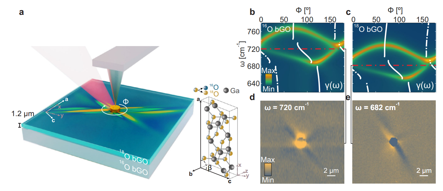
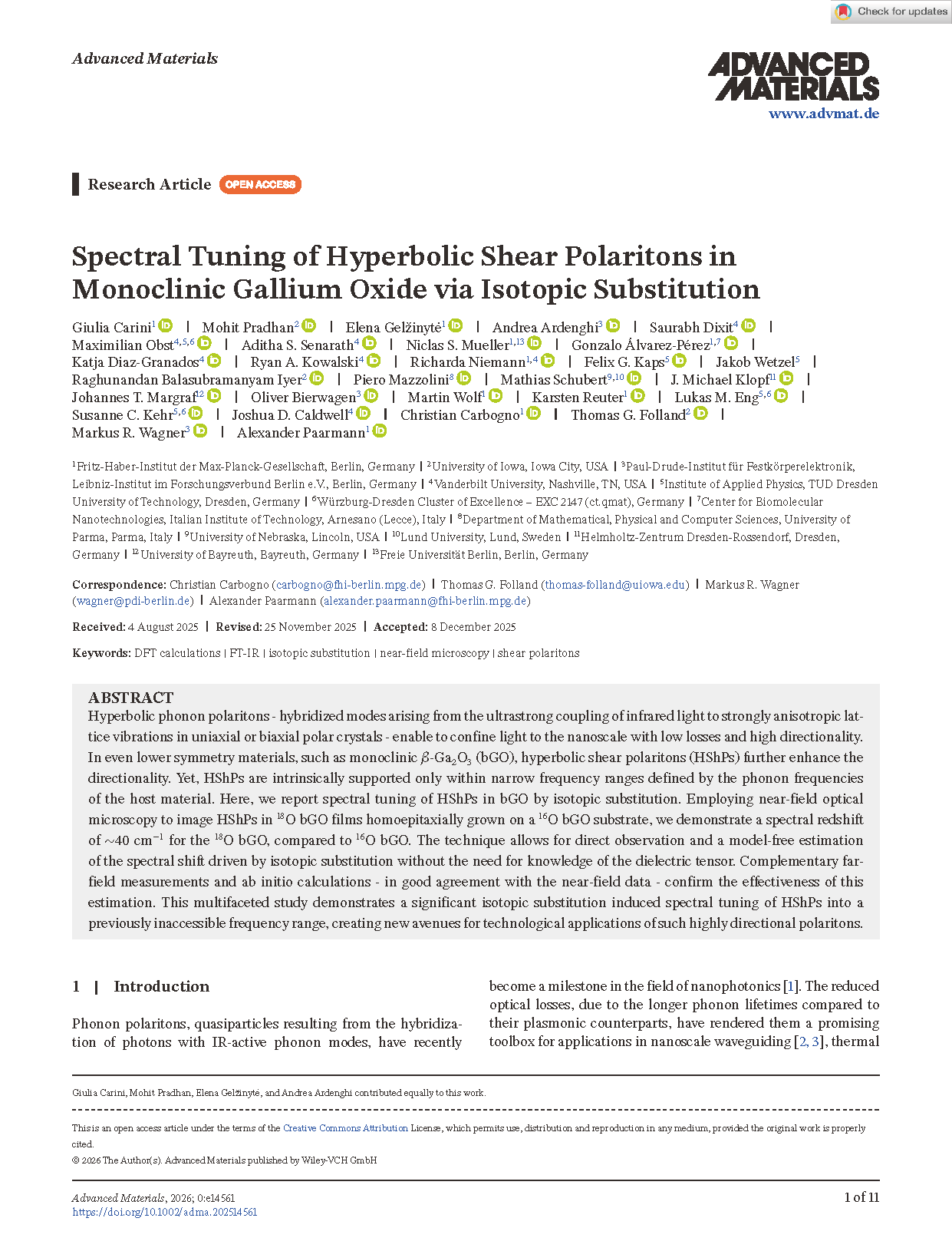
FIGURE 1 | Isotopic substitution-induced spectral shift of the hyperbolic shear polaritons in β-Ga₂O₃. (a) 3D schematic of the Free-Electron Laser (FEL)-coupled s-SNOM setup, illustrating the launching of HShPs by a Au disc on the surface of a 1.2 µm thick 18O bGO film homoepitaxially grown on a 16O bGO substrate. The inset shows a stick-and-ball representation of the conventional unit cell of bGO. The lattice constants were determined as a = 12.23 Å, b = 3.04 Å, and c = 5.80 Å, with the monoclinic angle between the a and c crystallographic axes being β = 103.76° [80]. The monoclinic a–c plane coincides with the sample surface. (b, c) Azimuthal dispersion for the 16O (b) and 18O (c) bGO isotopes obtained from transfer matrix simulations at a fixed in-plane momentum kip∕k0 = 1.1. The white curves indicate the axial dispersion, i.e., the frequency dependence of the optical axis direction γ(ω), calculated using Equation 1 (see Supporting Information, Section S7) with parameters derived from ab initio theory (Supporting Information, Tables S1 and S2). The red dashed-dotted lines indicate the frequencies at which the s-SNOM images in panels (d, e) were acquired. (d, e) Experimental near-field images of HShPs for the 16O (d) and 18O (e) bGO isotopes, taken at incident frequencies of 720 cm⁻¹ and 682 cm⁻¹, respectively.
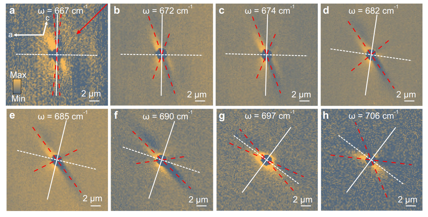
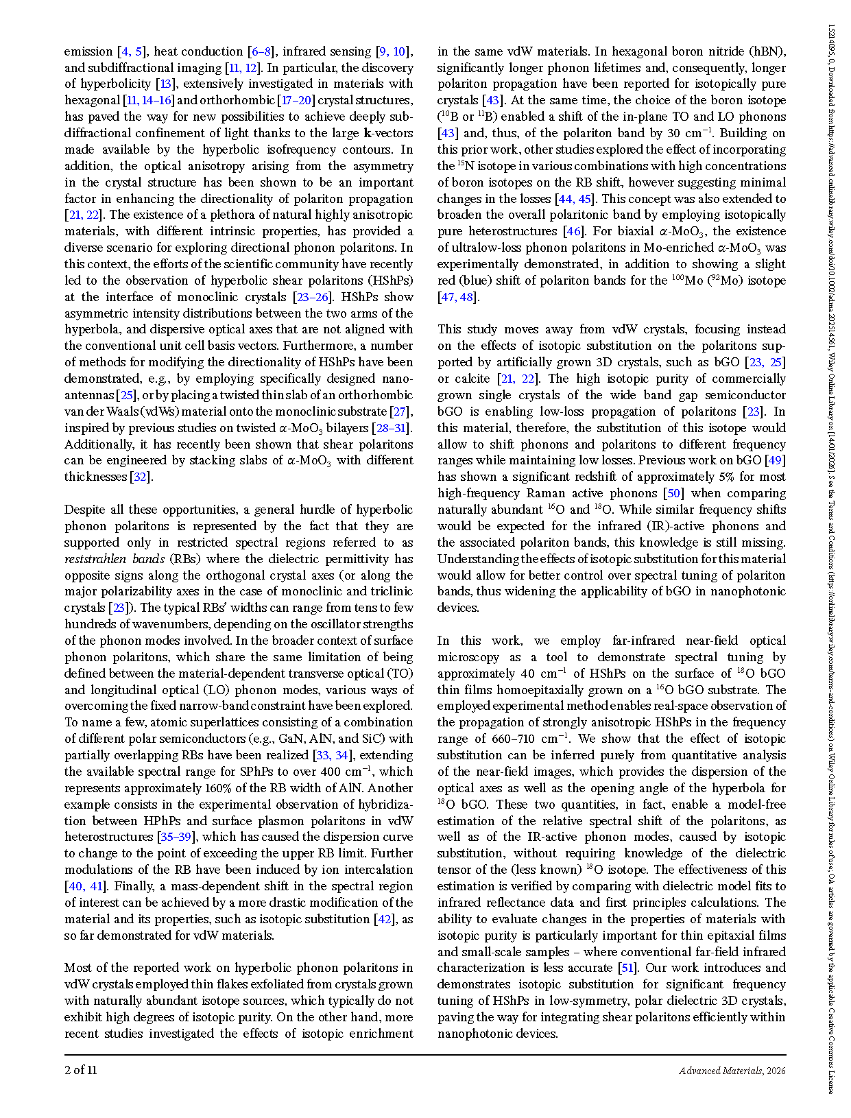
FIGURE 2 | Near-field propagation of low-symmetry hyperbolic polaritons in isotopically substituted 18O bGO film on a 16O bGO substrate. (a–h) Near-field microscopic images of HShPs in a 1.2 µm thick isotopically substituted bGO film homoepitaxially grown on a 16O bGO substrate. The modes are excited via a Au disk with a 2 µm diameter. The panels show images recorded at different excitation frequencies. For all images, the optical amplitude demodulated at the second harmonic of the tip tapping frequency (O2A) is displayed. The signal was acquired using a self-homodyne detection scheme. The red dashed lines indicate the asymptotes of the polariton wave fronts (see Supporting Information, Section S1 for details on their derivation). The white lines represent the two major polarizability axes of the material system. Panel (a) additionally provides information on the illumination direction (red arrow) and the crystallographic axes orientation (white arrows) (see Supporting Information).
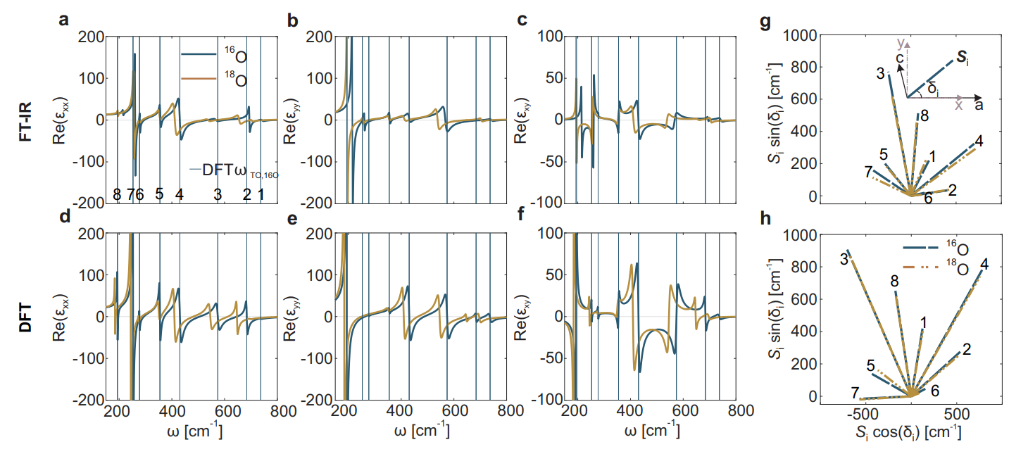
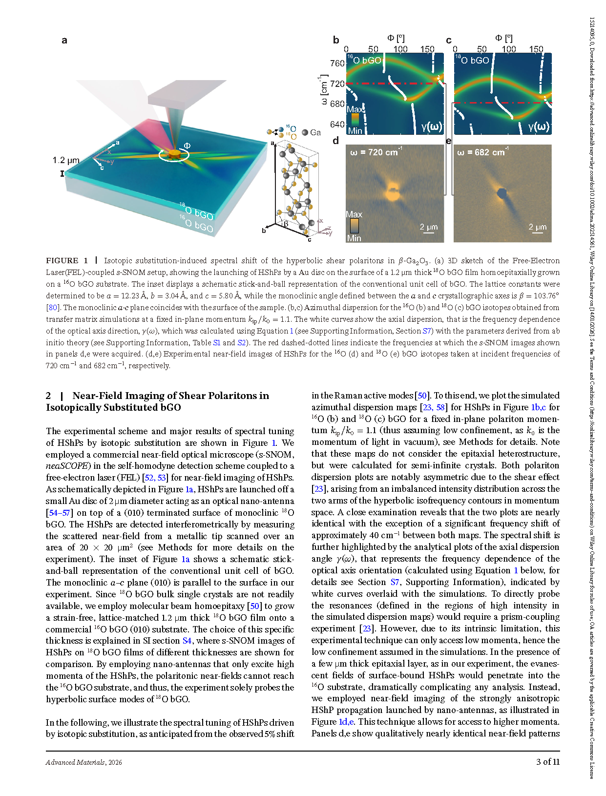
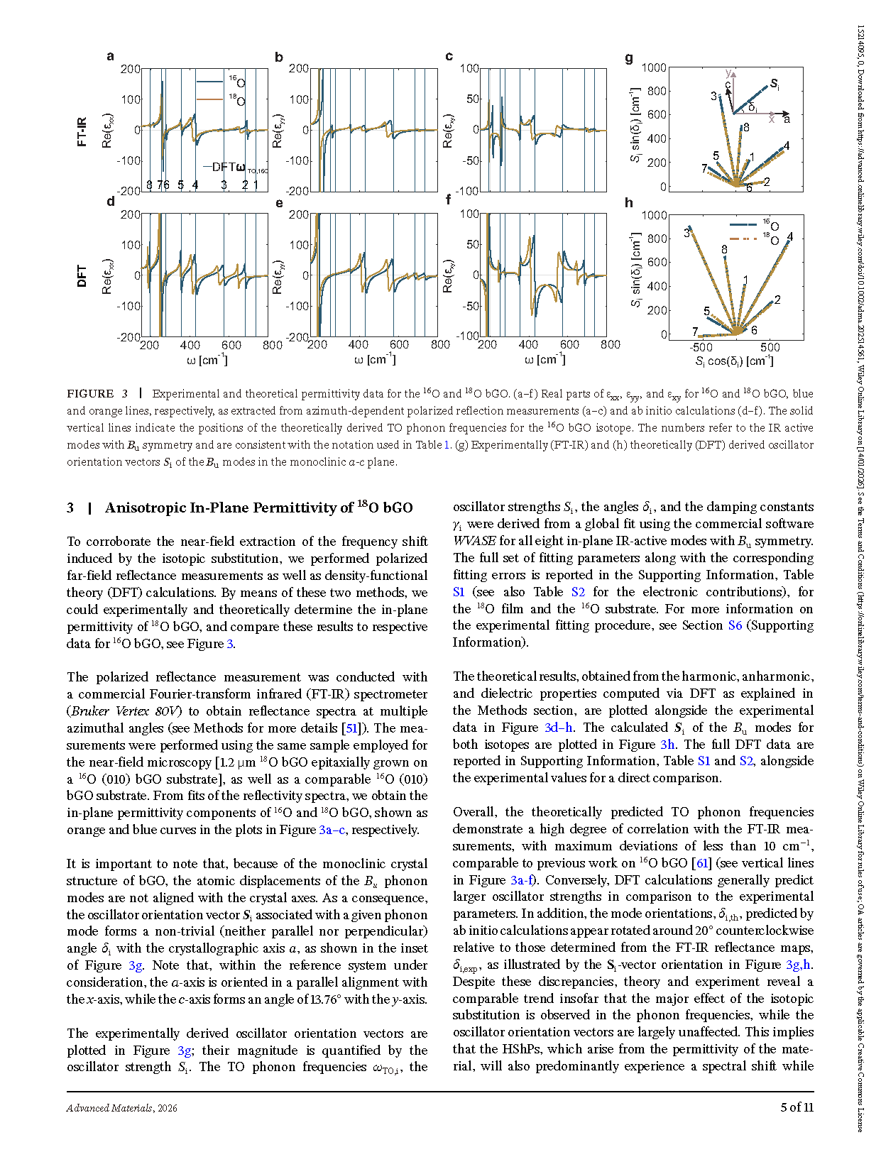
FIGURE 3 Experimental and theoretical permittivity data for the 16O and 18O bGO. (a–f) Real parts of ɛxx, ɛyy, and ɛxy for 16O and 18O bGO, blue and orange lines, respectively, as extracted from azimuth-dependent polarized reflection measurements (a–c) and ab initio calculations (d–f). The solid vertical lines indicate the positions of the theoretically derived TO phonon frequencies for the 16O bGO isotope. (g) Experimentally (FT-IR) and (h) theoretically (DFT) derived oscillator orientation vectors si of the Bu modes in the monoclinic a-c plane.
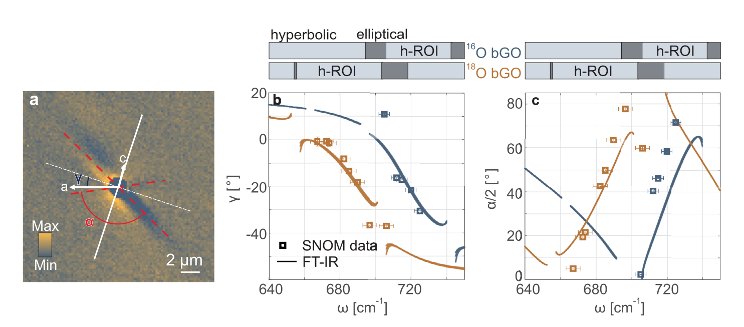
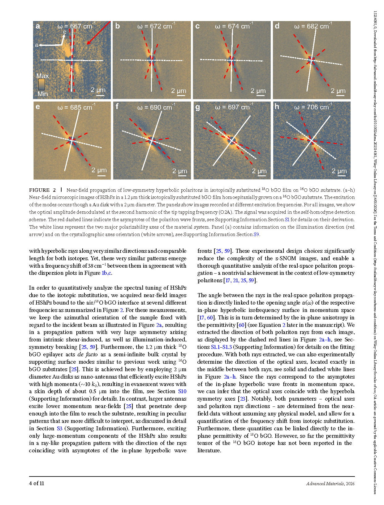
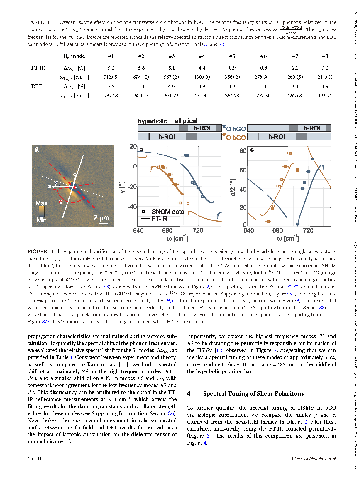
FIGURE 4 Experimental verification of the spectral tuning of the optical axis dispersion γ and the hyperbola opening angle a by isotopic substitution. (a) Illustrative sketch of the angles γ and a. While γ is defined between the crystallographic a-axis and the major polarizability axis (white dashed line), the opening angle a is defined between the two polariton rays (red dashed lines). As an illustrative example, we have chosen a s-SNOM image for an incident frequency of 690 cm−1 . (b,c) Optical axis dispersion angle γ (b) and opening angle a (c) for the 16O (blue curve) and 18O (orange curve) isotopes of bGO.
DOI:
doi.org/10.1002/adma.202514561