


Researchers from Fudan University, Suzhou Institute of Nano-Tech and Nano-Bionics, Chinese Academy of Sciences, Aerospace Information Technology University, Northwestern Polytechnical University have published a dissertation titled "Beyond Photoreceptors: Bio-Inspired Neuromorphic Vision System Mimicking Drosophila's Downstream Neural Circuitry With 2D β-Ga₂O₃ Schottky Diodes" in Rare Metals.
Background
Bio-inspired vision chips are core hardware for autonomous robots, visual prostheses and neuromorphic computing systems. Most existing optoelectronic devices only imitate photoreceptor photoelectric conversion, ignoring downstream neural processing units such as L1/L2 lamina cells in Drosophila, failing to realize on-chip complex visual computation including ON/OFF signal separation, temporal filtering and motion correlation multiplication, which creates a functional gap in hardware implementation. Two-dimensional β-Ga₂O₃ ultra-wide bandgap semiconductor owns atomically smooth dangling-bond-free surface, which enables near-ideal Schottky contacts. However, previous 2D Ga₂O₃ devices are only applied to UV detection rather than bionic motion sensing circuits. The classic Hassenstein-Reichardt motion detection model requires half-wave rectification and time-differential operation, while traditional silicon circuits suffer large footprint and poor high-frequency performance. No lightweight hardware solution based on 2D wide-bandgap diodes has been reported, and most works only verify simple grating detection without simulation of complex rotational motion, forming an obvious research gap.
Abstract
Bio‐inspired artificial vision systems have made significant progress, but most focus on photo‐receptors, neglecting the hardware implementation of the efficient downstream neural circuits for complex computations like motion detection. To overcome this bottleneck, we demonstrate a neuromorphic vision system that mimics Drosophila's motion detection pathway using high‐performance Schottky barrier diodes (SBDs) based on two‐dimensional β-Ga₂O₃ single crystals. The β-Ga₂O₃ SBDs exhibit an exceptional rectification ratio (>10⁸), a near‐ideal ideality factor of 1.08, and robust half‐wave rectification up to 10 MHz, enabled by an atomically sharp semiconductor‐metal interface. These properties replicate the Drosophila's motion detection circuitry: half‐wave rectification separates ON/OFF signals, whereas integrated resistor–capacitance (RC) circuits perform asymmetric temporal filtering. By implementing a hardware‐based Hassenstein‐Reichardt correlator model, our system successfully discriminates moving grating directions. Simulations of an 80 × 80 SBD array further demonstrate the decoding of complex rotational motion, generating optical flow fields, and identifying rotation centers. This work merges materials science and neuroscience through a hardware prototype that implements a complete bio‐inspired visual processing pipeline, paving the way for next‐generation compact intelligent vision systems.
Highlights
Propose a full hardware neuromorphic vision system beyond photoreceptors to realize Drosophila complete downstream motion neural computation.
Fabricate 2D single-crystal β-Ga₂O₃ SBD with atomically abrupt interface, rectification ratio >10⁸ and stable rectification up to 10 MHz.
Hardware implementation of Hassenstein‐Reichardt correlator to distinguish left/right moving grating signals.
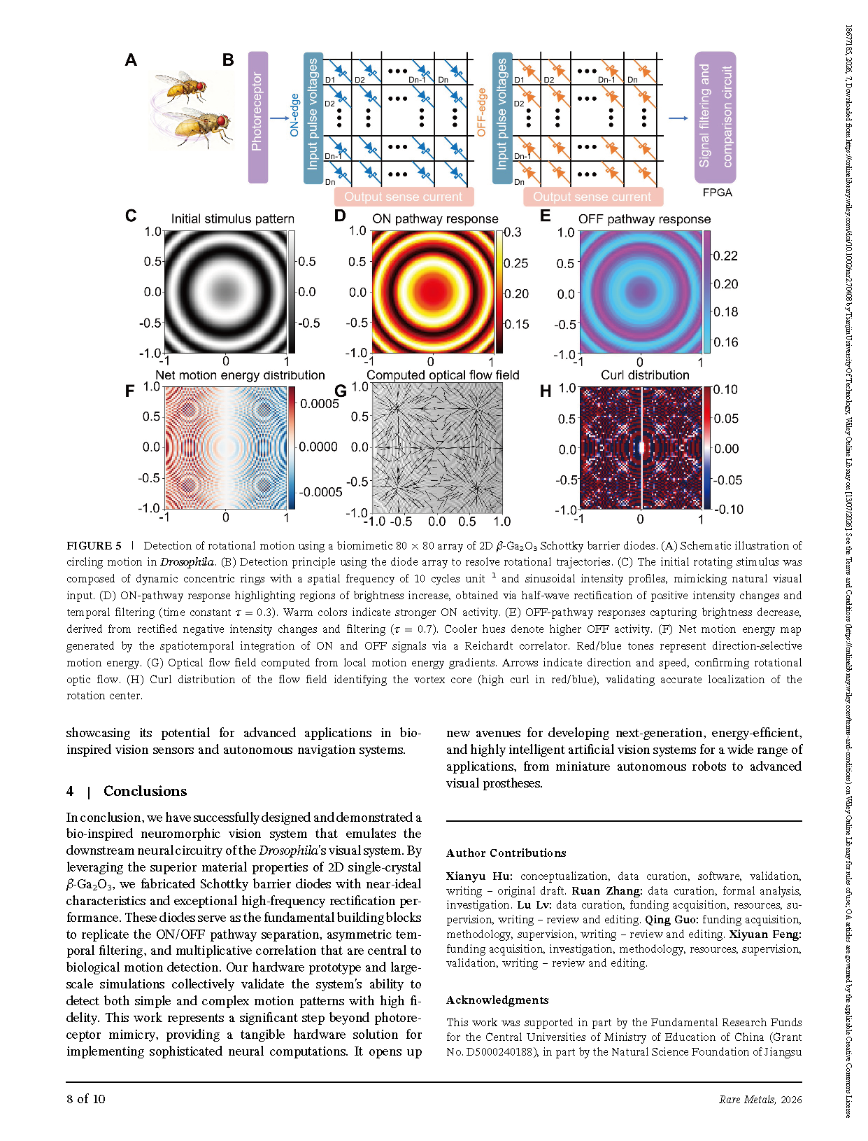
Complete 80 × 80 diode array simulation for rotational motion decoding, optical flow extraction and rotation center positioning.
Conclusion
In conclusion, we have successfully designed and demonstrated a bio‐inspired neuromorphic vision system that emulates the downstream neural circuitry of the Drosophila's visual system. By leveraging the superior material properties of 2D single‐crystal β-Ga₂O₃, we fabricated Schottky barrier diodes with near‐ideal characteristics and exceptional high‐frequency rectification performance. These diodes serve as the fundamental building blocks to replicate the ON/OFF pathway separation, asymmetric temporal filtering, and multiplicative correlation that are central to biological motion detection. Our hardware prototype and large‐scale simulations collectively validate the system's ability to detect both simple and complex motion patterns with high fidelity. This work represents a significant step beyond photoreceptor mimicry, providing a tangible hardware solution for implementing sophisticated neural computations. It opens up new avenues for developing next‐generation, energy‐efficient and highly intelligent artificial vision systems for a wide range of applications, from miniature autonomous robots to advanced visual prostheses.
Project Support
This work was supported in part by the Fundamental Research Funds for the Central Universities of Ministry of Education of China (Grant No. D5000240188), in part by the Natural Science Foundation of Jiangsu Province (Grant No. BK20240484), and in part by the Natural Science Foundation of Shandong Province (Grant No. ZR2025QC1603).
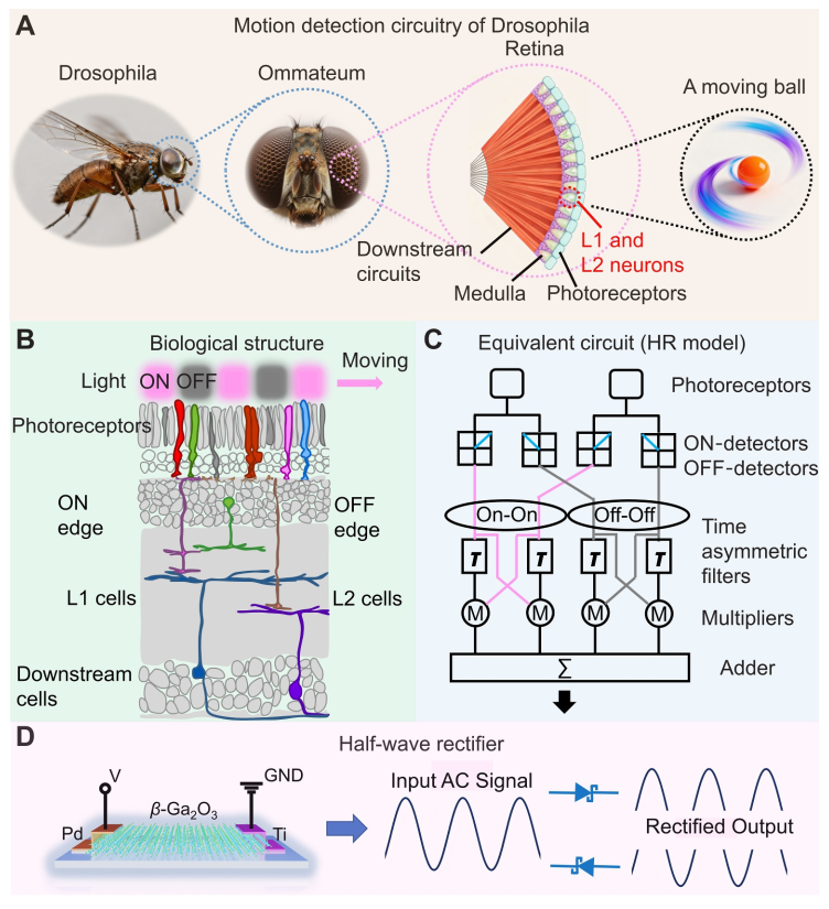
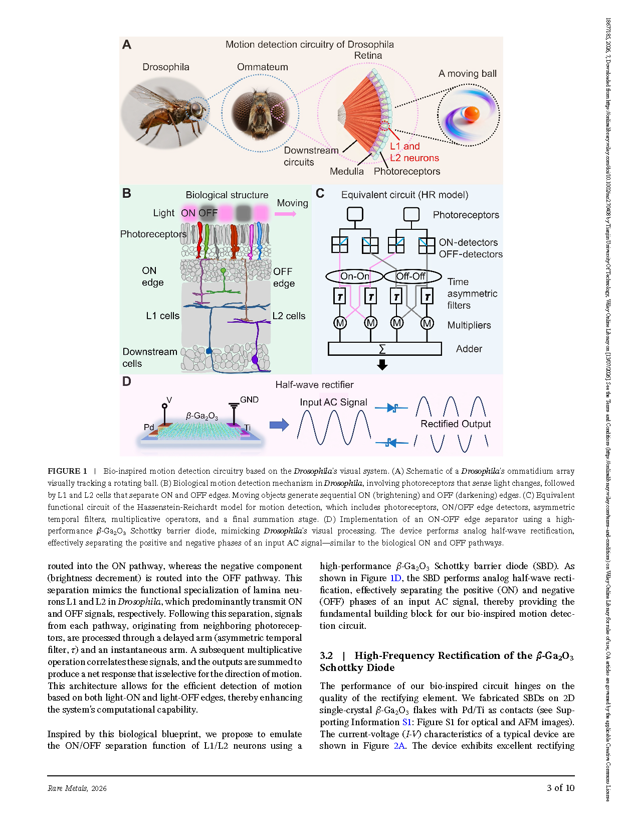
FIGURE 1 | Bio‐inspired motion detection circuitry based on the Drosophila's visual system. (A) Schematic of a Drosophila's ommatidium array visually tracking a rotating ball. (B) Biological motion detection mechanism in Drosophila, involving photoreceptors that sense light changes, followed by L1 and L2 cells that separate ON and OFF edges. Moving objects generate sequential ON (brightening) and OFF (darkening) edges. (C) Equivalent functional circuit of the Hassenstein‐Reichardt model for motion detection, which includes photoreceptors, ON/OFF edge detectors, asymmetric temporal filters, multiplicative operators, and a final summation stage. (D) Implementation of an ON‐OFF edge separator using a high‐performance β-Ga₂O₃ Schottky barrier diode, mimicking Drosophila's visual processing. The device performs analog half‐wave rectification, effectively separating the positive and negative phases of an input AC signal—similar to the biological ON and OFF pathways.
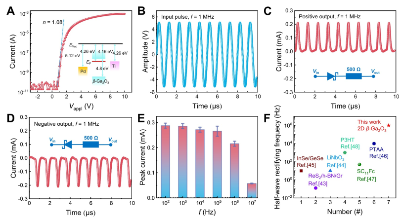
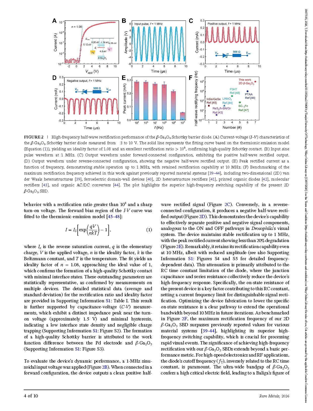
FIGURE 2 | High‐frequency half‐wave rectification performance of the β-Ga₂O₃ Schottky barrier diode. (A) Current‐voltage (I–V) characteristics of β-Ga₂O₃ Schottky barrier diode measured from −3 to 10 V. The solid line represents the fitting curve based on the thermionic emission model (Equation (1)), yielding an ideality factor of 1.08 and an excellent rectification ratio ratio >10⁸, confirming high‐quality Schottky contact. (B) Input sine pulse waveform at 1 MHz. (C) Output waveform under forward‐connected configuration, exhibiting the positive half‐wave rectified output. (D) Output waveform under reverse‐connected configuration, showing the negative half‐wave rectified output. (E) Peak rectified current as a function of frequency, demonstrating stable operation up to 1 MHz, with retained rectification capability at 10 MHz. (F) Benchmarking of the maximum rectification frequency achieved in this work against previously reported material systems, including two‐dimensional (2D) van der Waals heterostructures, ferroelectric domain‐wall devices, 2D heterostructure rectifiers, printed organic diodes, molecular rectifiers , and organic AC/DC converters. The plot highlights the superior high‐frequency switching capability of the present 2D β-Ga₂O₃ SBD.
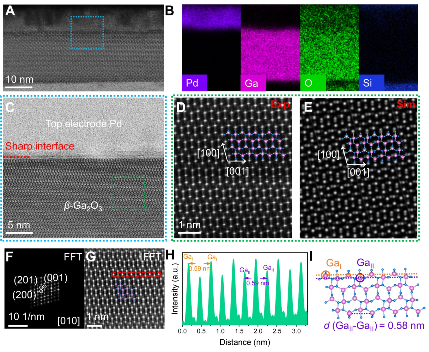
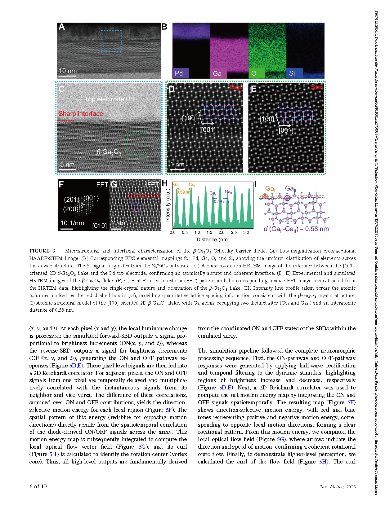
FIGURE 3 | Microstructural and interfacial characterization of the β-Ga₂O₃ Schottky barrier diode. (A) Low‐magnification cross‐sectional HAADF‐STEM image. (B) Corresponding EDS elemental mappings for Pd, Ga, O, and Si, showing the uniform distribution of elements across the device structure. The Si signal originates from the Si/SiO₂ substrate. (C) Atomic‐resolution HRTEM image of the interface between the [100]‐ oriented 2D β-Ga₂O₃ flake and the Pd top electrode, confirming an atomically abrupt and coherent interface. (D, E) Experimental and simulated HRTEM images of the β-Ga₂O₃ flake. (F, G) Fast Fourier transform (FFT) pattern and the corresponding inverse FFT image reconstructed from the HRTEM data, highlighting the single‐crystal nature and orientation of the β-Ga₂O₃ flake. (H) Intensity line profile taken across the atomic columns marked by the red dashed box in (G), providing quantitative lattice spacing information consistent with the β-Ga₂O₃ crystal structure. (I) Atomic structural model of the [100]‐oriented 2D β-Ga₂O₃ flake, with Ga atoms occupying two distinct sites (GaⅠ and GaⅡ) and an interatomic distance of 0.58 nm.
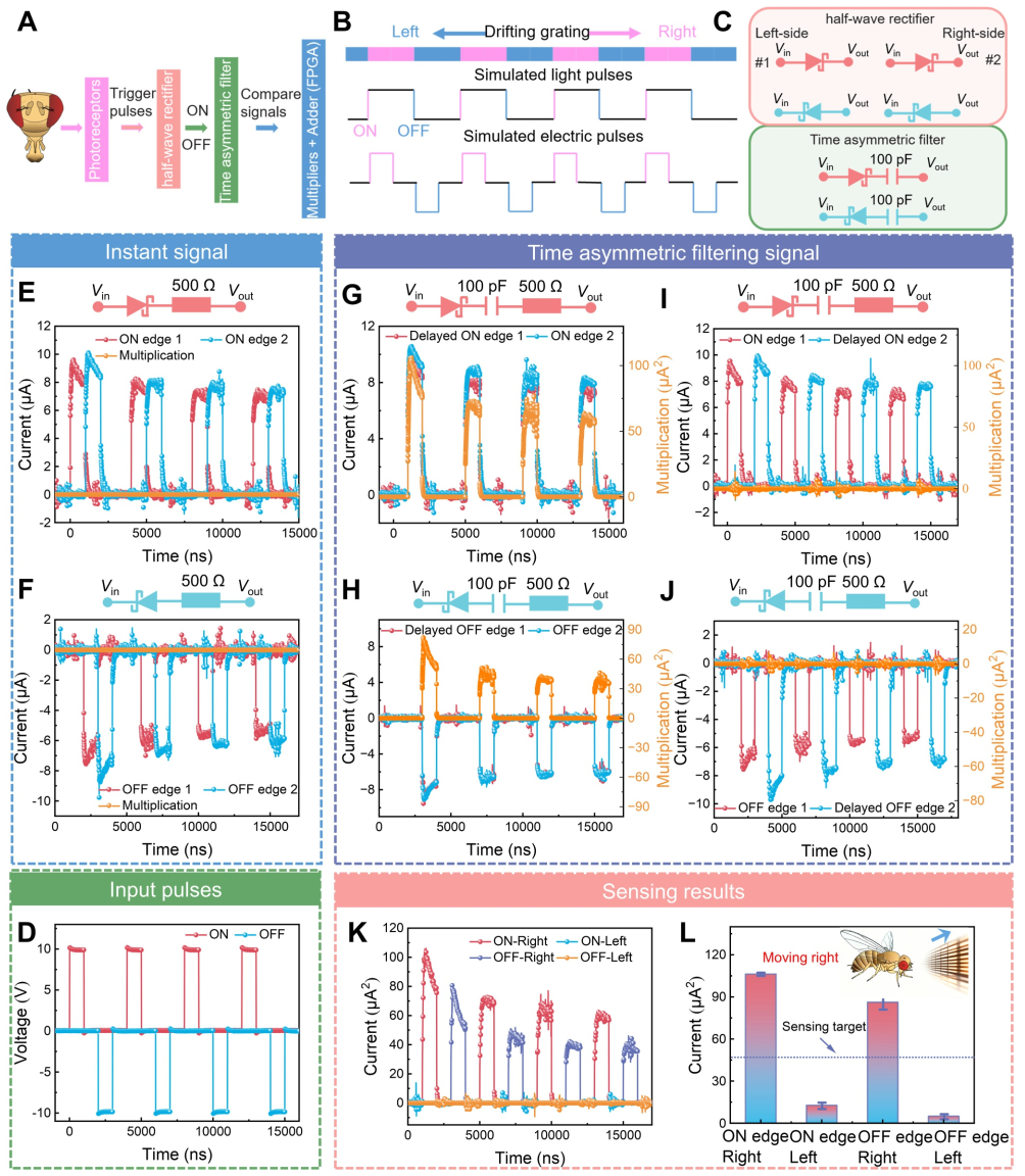
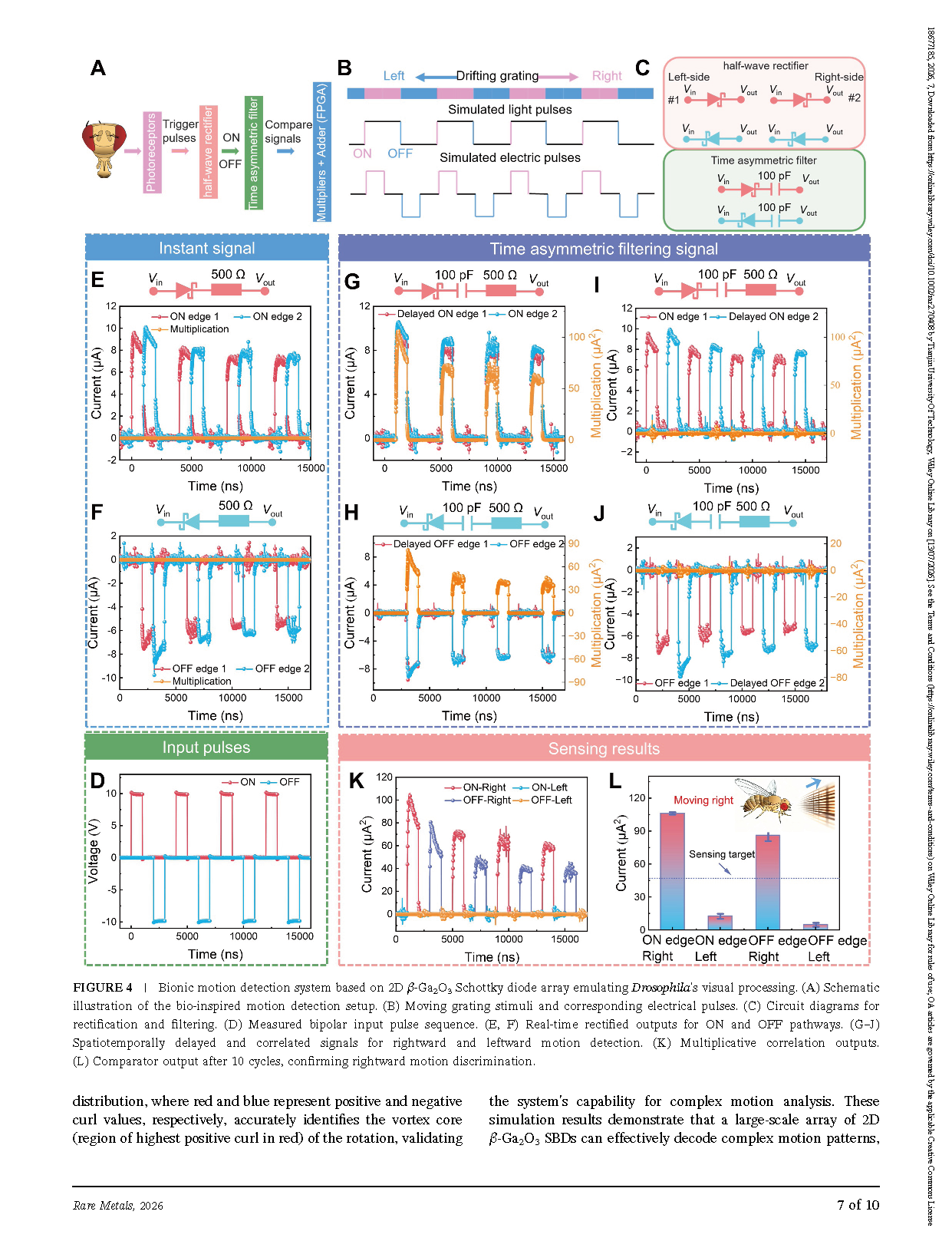
FIGURE 4 | Bionic motion detection system based on 2D β-Ga₂O₃ Schottky diode array emulating Drosophila's visual processing. (A) Schematic illustration of the bio‐inspired motion detection setup. (B) Moving grating stimuli and corresponding electrical pulses. (C) Circuit diagrams for rectification and filtering. (D) Measured bipolar input pulse sequence. (E, F) Real‐time rectified outputs for ON and OFF pathways. (G–J) Spatiotemporally delayed and correlated signals for rightward and leftward motion detection. (K) Multiplicative correlation outputs. (L) Comparator output after 10 cycles, confirming rightward motion discrimination.
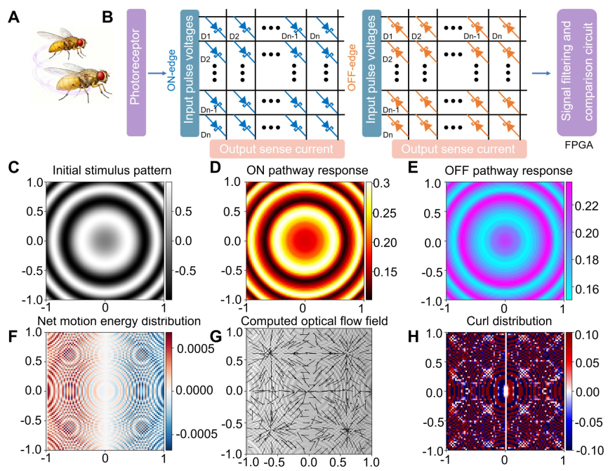
FIGURE 5 | Detection of rotational motion using a biomimetic 80 x 80 array of 2D β-Ga₂O₃ Schottky barrier diodes. (A) Schematic illustration of circling motion in Drosophila. (B) Detection principle using the diode array to resolve rotational trajectories. (C) The initial rotating stimulus composed of dynamic concentric rings with a spatial frequency of 10 cycles unit⁻¹ and sinusoidal intensity profiles, mimicking natural visual input. (D) ON‐pathway response highlighting regions of brightness increase, obtained via half‐wave rectification of positive intensity changes and temporal filtering (time constant τ=0.3). Warm colors indicate stronger ON activity. (E) OFF‐pathway responses capturing brightness decrease, derived from rectified negative intensity changes and filtering (τ=0.7). Cooler hues denote higher OFF activity. (F) Net motion energy map generated by the spatiotemporal integration of ON and OFF signals via a Reichardt correlator. Red/blue tones represent direction‐selective motion energy. (G) Optical flow field computed from local motion energy gradients. Arrows indicate direction and speed, confirming rotational optic flow. (H) Curl distribution of the flow field identifying the vortex core (high curl in red/blue), validating accurate localization of the rotation center.
DOI:
doi.org/10.1002/rar2.70408
Instruction
Official Account

Cooperation
Top